Anatomy of the Bowman’s Capsule
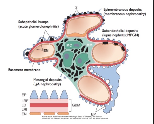
EP : epithelial membrane
LRE : Lamina rara externa
LD : Lamina densa
LRI : Lamina rara interna
EN : Endothelial membrane
The Glomerular Basement Membrane
| Layer | Location | Composition | Function |
| lamina rara externa | adjacent to podocyte processes | heparan sulfate | blocks by charge |
| lamina densa | dark central zone | type 4 collagen and laminin | blocks by size (Molecular Weight > 5800kDa) |
| lamina rara interna | adjacent to endothelial cells | heparan sulfate | blocks by charge |
Linear IgG deposition, occurs in:
- Anti GBM disease (crescents, very strong staining)
- Diabetes (no crescents, diabetic glomerosclerosis, IgG is non selectively absorbed into the highly permeable capillary wall, there is deposition of albumin & other plasma proteins)
- Fibrillary GN (IgG absorbed into the fibrils)
- Light chain disease
- Alport’s after transplantation
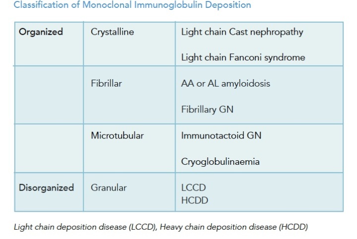
Nodular mesangium:
- Light chains
- Amyloid
- Diabetes
- Chronic MPGN
- Organised glomerular deposits disease
- fibrillary GN
- immunotactoid GN
- fibronectin GN
- collagen III GN
- Idiopathic
- Smoking
- HTN
- Chronic hypoxic/ischaemic conditions
- Takayasu’s , RAS
- cyanotic congenital heart disease
- CF
Granular IgG deposition:
- membranous
- MPGN
- PIGN
Mesangial Proliferation Differential Diagnosis:
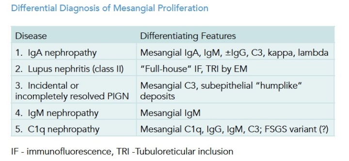
EM
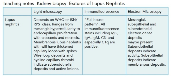
Presence of tubuloreticular inclusion bodies:
- lupus nephritis
- Alfa interferon therapy
- HIV(AN)
- Viral infections
Full house Ig staining:
- Lupus
- HIVICK
- SBE / shunt nephritis
- PIGN (Rare)
Anti Ro/SSA Ab : Associated with SLE/PBC
Anti La/SSB Ab : Sjogrens
Anti – RNP Ab : MCTD/SLE. More prominent in Raynaud’s with mild renal involvment
Anti Scl-70 Ab : Systemic sclerosis
Anti Sm Ab : SLE. Assoc with increased severity and activity of renal disease. Can be induced by EBV by molecular mimicry.
Anti Mi-2 : 25% of dermatomyositis
Anti Jo-1 : Polymyositis (+/- dermatomyositis)
Appearances of monoclonal immunoglobulin deposition disease